2. EEG artifact removal using Blind Source Separation
Severe contamination of EEG activity by eye movements, blinks, muscle, heart and line noise is a serious problem for EEG interpretation and analysis. Many methods have been proposed to remove eye movement and blink artifacts from EEG recordings:
-
•Simply rejecting contaminated EEG epochs results in a considerable loss of collected information.
-
•Often regression in the time or frequency domain is performed on simultaneous EEG and electrooculographic (EOG) recordings to derive parameters characterizing the appearance and spread of EOG artifacts in the EEG channels. However, EOG records also contain brain signals, so regressing out EOG activity inevitably involves subtracting a portion of the relevant EEG signal from each recording as well.
-
•Since many noise sources, include muscle noise, electrode noise and line noise, have no clear reference channels, regression methods cannot be used to removed them.
We propose to apply ICA to multichannel EEG recordings and remove a wide variety of artifacts from EEG records by eliminating the contributions of artifactual sources onto the scalp sensors. Our results show that ICA can effectively detect, separate and remove activity in EEG records from a wide variety of artifactual sources, with results comparing favorably to those obtained using regression-based and Principal Component Analysis methods.
ICA Assumptions
ICA-based artifact correction can separate and remove a wide variety of artifacts from EEG data by linear decomposition. The ICA method is based on the assumptions that the time series recorded on the scalp:
-
•are spatially stable mixtures of the activities of temporally independent cerebral and artifactual sources, that
-
•the summation of potentials arising from different parts of the brain, scalp, and body is linear at the electrodes, and that
-
•propagation delays from the sources to the electrodes are negligible.
Assumptions two and three above are quite reasonable for EEG (or MEG) data. Given enough input data, the first assumption is reasonable as well. The method uses spatial filters derived by the ICA algorithm, and does not require a reference channel for each artifact source. Once the independent time courses of different brain and artifact sources are extracted from the data, artifact-corrected EEG signals can be derived by eliminating the contributions of the artifactual sources.
Methods
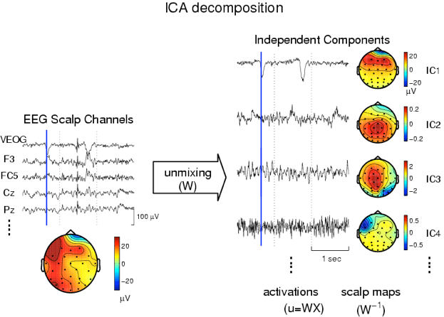
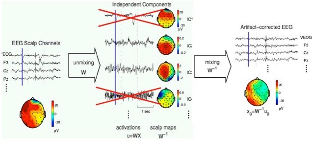
The figure below presents a schematic illustration of the procedure (Click on figure to expand). In EEG analysis, the rows of the input matrix, X, are EEG signals recorded at different electrodes and the columns are measurements recorded at different time points (left). ICA finds an `unmixing' matrix, W, which decomposes or linearly unmixes the multi-channel scalp data into a sum of temporally independent and spatially fixed components. The rows of the output data matrix, U = WX, are time courses of activation of the ICA components. The columns of the inverse matrix, inv(W), give the relative projection strengths of the respective components at each of the scalp sensors (right). These scalp weights give the scalp topography of each component, and provide evidence for the components' physiological origins. For instance:
Some Useful Heuristics
-
•Eye movements should project mainly to frontal sites with a lowpass time course.
-
•Eye blinks should project to frontal sites and have large punctate activations.
-
Temporal muscle activity should project to temporal sites with a spectral peak above 20 Hz.
Component Projections
Calling the activations the matrix of unmixed component time courses,
W = weights * sphere;
-
activations = W * data;
and the inverse weight matrix (i.e., the mixing matrix),
Winv = inv(W);
(or Winv = pinv(W); if the number of components is les than the number of channels), then the projection of the i'th independent component onto the original data channels is given by
projection = Winv(:,i) * activations(i,:);
The projection of the i'th component is the outer product of i'th row of the component activation (i.e., the component time course), activations(i,:), with the i'th column of the inverse matrix (i.e., the component scalp map), Winv(:,i). The projected component data has the same size as the original data, has the same basis (i.e. each row is a single electrode, as in the original data), and is scaled in the original data units (e.g., uV). Scaling information and polarity are distributed between the activation waveforms and the maps. This means the true size (and polarity) of a component is given by the size (and polarity) of its projection.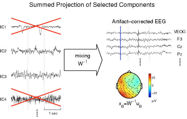
For the data shown above, all scalp maps were interpolated from 31 EEG channels and referred to the original right-mastoid reference. For each component, the amplitudes of scalp maps (given by the individually scaled color bars of the right panel) give the size of the component projections at the time point marked by the vertical blue line.
Above, artifact-free event-related brain signals were obtained by projecting the sum of selected non-artifactual ICA components back onto the scalp,
clean_data = Winv(:,a) * activations(a,:);
where [a] was a vector of the selected non-artifactual component numbers. The toolbox contains a function, icaproj(), that computes projections in a single line of code.
clean_data = icaproj(data,W,a);
In practice, the trick is to decide which components, [a], are artifactual. Above, we list some heuristics we have found useful.
Sample Experimental Data -
Removing blink and muscle artifacts
The figure below shows a 3-sec portion of the recorded EEG time series and its ICA component activations, the scalp topographies of four selected components, and the artifact-corrected EEG signals obtained by removing four selected EOG and muscle noise components from the data. The eye movement artifact at 1.8 sec in the EEG data (left) is isolated to ICA components 1 and 2 (left middle). The scalp maps (right middle) indicate that these two components account for the spread of EOG activity to frontal sites.
Eliminating the four artifact components whose scalp maps are shown above, and projecting the remaining components back onto the scalp channels produced artifact-corrected EEG data (right) free of these artifacts.
Note that removing the eye blink activity from frontal channels (Fp1, Fp2 left panel) clearly reveals frontal alpha activity occurring during the blink that is obscure in the original data.
Note also the regular right fronto-temporal muscle spike component #13 (middle panel) which, though difficult to see in the original data (e.g., in channel: T4), was nevertheless cleanly separated from other activity by ICA. -
Some heavily contaminated EEG data
A five-second portion of a corrupted EEG time series resulting from a poor data-acquisition setting; (B) noise components extracted by ICA (right panel). (C) The same EEG signals corrected for artifacts by ICA by removing the six selected components, and, (D) spectral analysis of the original and artifact-corrected EEG recordings. Note that EEG activity is more visible than in (A), particularly in channels 1 and 2, and the line noise (60 Hz) and aliased line noise frequencies (near 12 Hz, 105 Hz, 135 Hz) are reduced.
References
Our approach to artifact correction using ICA is available in two journal articles:
-
-
•Jung T-P, Makeig S, Humphries C , Lee TW, McKeown MJ, Iragui V, and Sejnowski TJ, "Removing Electroencephalographic Artifacts by Blind Source Separation," Psychophysiology, 37:163-78, 2000 (.pdf, 1.3Mb).
-
•Jung T-P, Makeig S, Westerfield W, Townsend J, Courchesne E, and Sejnowski TJ, "Removal of eye activity artifacts from visual event-related potentials in normal and clinical subjects," Clinical Neurophysiology 111:1745-58, 2000 (.pdf, 4.9Mb).
-
-
-
•Other relevant references:
-
•Makeig S, Bell AJ, Jung T-P, and Sejnowski TJ, "Independent component analysis of Electroencephalographic data." Advances in Neural Information Processing Systems 8, 145-151,1996.
-
•Jung T-P, Humphries C, Lee TW, Makeig S, McKeown MJ, Iragui V, and Sejnowski TJ, "Extended ICA Removes Artifacts from Electroencephalographic Recordings", Advances in Neural Information Processing Systems 10:894-900, 1998.
-
•Jung T-P, Humphries C, Lee TW, Makeig S, McKeown MJ, Iragui V, and Sejnowski TJ, "Removing Electroencephalographic Artifacts : Comparison between ICA and PCA", In: Neural Networks for Signal Processing VIII, 63-72, 1998.
-
•Jung T-P, Makeig S, Westerfield M, Townsend J, Courchesne E, and Sejnowski TJ, "Analyzing and Visualizing Single-trial Event-related Potentials," In: Advances in Neural Information Processing Systems, 11:118-24, 1999.
-
Visit the ICA Toolbox tutorial
Download the ICA toolbox
This section on artifact rejection by
Tzyy-Ping Jung & Scott Makeig jung@sccn.ucsd.edu
We welcome comments and suggestions. scott@sccn.ucsd.edu
ICA separates underlying brain and artifactual sources.
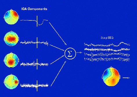
EEG artifact removal with Blind Source Separation