SINGLE-TRIAL ERPS DURING CONTINUOUS fMRI SCANNING.
T-P. Jung, S. Makeig, M. Westerfield, B. Hicks, E. Courchesne and T. J. Sejnowski. Children's Hospital Research Center, Naval Health Research Center, Howard Hughes Medical Institute and Salk Institute, Institute for Neural Computation, University of California San Diego
Electromagnetic and functional magnetic resonance brain imaging measures have complementary strengths and weaknesses in temporal and spatial resolutions. First approaches to combining electrophysiological and hemodynamic brain data have compared the scalp topography of event-related potential (ERP) averages against task-related fMRI activation level differences (George et al., 1995; Buckner et al., 1996; McCarthy et al., 1997) based on ERP and fMRI data collected in separate experimental sessions. Shortcomings of this approach include (1) difficulty in co-registration; (2) order effects in separately recorded data sets; (3) averaged ERP data reveal only a small portion of event-related brain dynamics (Makeig, 1993). We used a custom EEG amplifier with timeout circuits and shielded cabling (SA Instruments) to record single-trial event-related potentials (ERPs) at site C2 from a healthy adult subject who attended to shapes (nontarget-frequent circles and target-squares (25%) flashed on a projector screen during continuous 1.5T fMRI scanning.
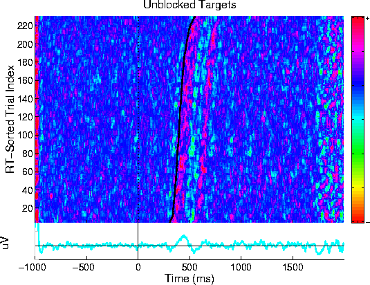
Target Stimulus-Aligned ERP Image This plot shows an "ERP image" plot of the 226 single trials (color-coded as single horizontal lines in the image) after blanking out of the pulse-artifact in each trial and removal of the mean pulse responses. The target response mean is shown in the light blue trace below the ERP image.
After time-limited artifact rejection, the `ERP-image' plot (Jung et al., 1999) showed clearly that the response-locked brain activity was precisely time locked to subject responses in nearly all trials, while the stimulus-locked ERP was time-locked to stimulus onsets. The average of the available ERPs included the well-documented P1/N1/P2/N2 and target LPC components. The fMRI data from this experiment showed no decrement in image quality from the EEG electrodes or recording equipment. Standard correlational analysis showed task-related activations in motor cortex and right anterior cerebellum, as would be expected in a right-handed subject.
Research supported by the Office of Naval Research, Howard Hughes Medical Institute, National Institutes of Health NS34155 and MH36840 and the Swartz Foundation.