Chapter_01_Getting_Started_with_NFT
The Neuroelectromagnetic Forward Head Modeling Toolbox is an open-source software toolbox running under MATLAB (The Mathworks, Inc.) for generating realistic head models from available data (MRI and/or electrode locations) and for solving the forward problem of electro-magnetic source imaging. The toolbox includes tools for segmenting scalp, skull, cerebrospinal fluid (CSF) and brain tissues from T1-weighted magnetic resonance (MR) images. After extracting the segmented tissue volumes, mesh generation can be performed. When MR images are not available, it is possible to warp a template head model to measured electrode locations to obtain a better-fitting head model. The toolbox also includes electrode scalp mesh co-registration and generation of a uniform source space inside the brain volume for to be used in coarse source localization. The Boundary Element Method (BEM) is used for the numerical solution of the forward problem. Toolbox functions can be called from either a graphic user interface or from the command line. Function help messages and a tutorial are included. The toolbox is freely available under the GNU Public License for noncommercial use and open source development.
The toolbox uses the following third party tools and libraries for segmentation, mesh generation and forward problem solution. The source codes for these tools are available.
1. ASC - for triangulation of 3D volumes.
2. Qslim - for mesh coarsening.
3. Matitk - Matlab interface to the ITK image processing toolkit.
4. Metu-bem - Boundary Element Method solver.
The NFT toolbox provides a user interface (UI) for segmentation, mesh generation and for creating the numerical head model. It also has a well defined MATLAB command-line interface.
This manual explains how to use the NFT toolbox. The head modeling UI, the command line API and the structures are described. An overview of the implementation is provided.
The next section describes the installation of the toolbox. The Getting Started section provides an overview of the interface. Head modeling from 3D MR images is described next, followed by head modeling from template warping. This is followed by a section on forward modeling and examples. The final section is a summary of all toolbox functions and commands.
This section describes installation and configuration of the NFT Toolbox. The following steps are necessary for a proper installation of the toolbox:
1. Extract or copy the toolbox directory to a suitable place on your computer file system.
2. The extracted directory will contain m-files, and C++ executables.
3. Add the toolbox directory to the MATLAB path. You can use the File → SetPath menu item or the addpath() function. Under linux/unix, you may add the directory to the MATLABPATH.
The toolbox can also make use of the Matlab Parallel Processing toolbox (if installed) to distribute the computation of the transfer and lead-field matrices to multiple processors. To do this, before running NFT, the user must simply enter
>> matlabpool(n) % where n is the number of compute nodes available
In parallel mode, wait bars do not appear while computing the transfer and lead-field matrices.
The toolbox starts by typing Neuroelectromagnetic_Forward_Modeling_Toolbox or NFT on command window. Main window appears as shown in Figure 1. This window is divided into three panels. The first panel is used to select the working folder, and to name the subject and the session. The NFM toolbox requires a subject folder to be specified at startup. All subject specific output is saved into this folder. The filenames are derived from the subject and session names entered into this panel. The second panel is the Head modeling panel. The head model can either be created from MR images, or a template head model can be warped to digitized sensors. The head modeling panel provides the following operations when creating a head model from MR images:
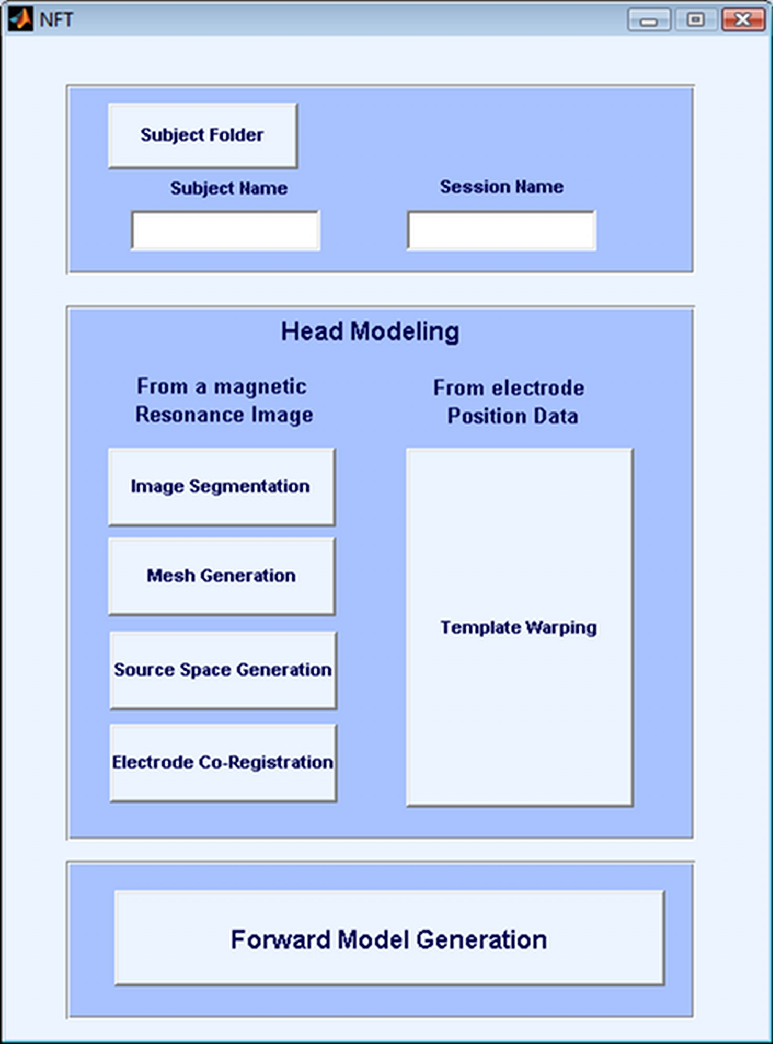
Image Segmentation
Interface for tissue classification from 3D MR Images.
Mesh Generation
Uses the segmentation results to generate realistic BEM meshes.
Source Space Generation
Generates a regular grid sources within the brain mesh.
Electrode Co-Registration
Registers digitized electrode locations to the scalp mesh.
When generating a template-based head model from digitized electrode positions, the only option is Template Warping. The final panel in the main menu is for Forward Model Generation. This opens up the Forward Model Generation interface which is used to compute the BEM coefficient matrix, create the transfer matrices for each sensor, and generate lead field matrices for a source distribution.