NFT_Appendix_C
An important consideration for obtaining accurate forward problem solutions is to correctly model the distribution of conductivity within the head. In the literature, consistent conductivity values have been reported for scalp, brain, and CSF tissues but there has been a huge variation in reported skull conductivity values. This is partly caused by variations in skull conductivity from person to person and throughout the life cycle (Hoekema et al 2003), and partly from use of different measurement/estimation methods (Oostendorp et al 2000). In the 1970's and 80's the brain-to-skull conductivity ratio was reported to be 80 (Rush 1968, Cohen 1983), still a very commonly used ratio in EEG source localization. More recent studies in the last decade have reported this ratio to be as low as 15 (Oostendorp 2000). In a more recent study on epilepsy patients undergoing presurgical evaluation using simultaneous intra-cranial and scalp EEG recordings, the average brain-to-skull conductivity ratio was estimated to be 25 (Lai 2005).
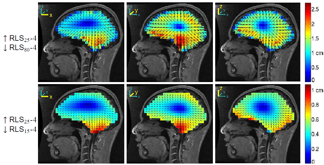
Below, we present some simulation results showing the effects of using incorrect skull conductivity values on equivalent dipole source localization. For this purpose, we solved the forward electrical head model problem using a realistic, subject-specific four-layer BEM model built from a subject’s MR head image using the NFT toolbox (Akalin Acar & Makeig, 2010). We set the forward model (‘ground truth’) brain-to-skull conductivity ratio to 25 and then solved the inverse problem using the same realistic head model using the commonly used ratio of 80. This produced equivalent dipole localization errors of up to 2.5 cm (figure below, top row). The positions of the grid of model dipoles were moved towards the scalp surface. On the other hand, (figure below, bottom row) if the brain-to-skull conductivity ratio was mis-estimated to be 15 when solving the inverse problem, the dipoles were localized more towards the center of the brain with localization errors up to 1 cm (Akalin Acar & Makeig, 2012). Therefore, correct modeling of skull conductivity is an important factor for EEG source localization.

Figure 1. Equivalent dipole source localization error directions (arrows) and magnitudes (colors) for a 4-layer realistic BEM head model when the brain-to-skull conductivity ratio was estimated to be 80 as opposed to the actual simulated forward model value of 25 (top row) and as 15 (as opposed to 25) (bottom row). The source space was a regular Cartesian grid of single equivalent dipole sources with 8-mm spacing filling the brain volume. The three columns show the errors when the equivalent dipole sources are oriented in x-, y-, and z-directions, respectively.