Advantage in accuracy for colored images decreases
for subject with fast behavioral responses
Categorization of familiar versus new
images in humans (behavior and EEG)
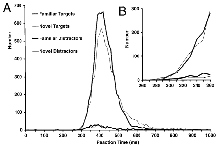
Human subjects had to categorize the same 200
images for 15 consecutive days. Then these images were mixed with 1200 previously
unseen ones to asses the difference between familiar and new images. Surprisingly,
for images categorized in under 250ms in humans they were no significant difference
between familiar and new images (below are the reaction times of all human
subjects, B is a zoom of fastest reaction times in A).

Moreover for
humans, early ERPs for familiar and new images were hardly distinguishable
(see figure below, on the left the grand average ERPs of 14 subjects and on
the right the differential ERPs for the two type of images). For more details, this work has been
pulished in Journal of Cognitive Neuroscience (see publication).

categorization versus
detection of target in natural images (EEG)
14 human subjects
alternated between two tasks. In one tasks, the subjects had to perform the
standard animal categorization task (see above). In the other tasks, subject
had to categorize a single image (that contained an animal for comparison
with the categorization task) among various non-target images (as in the
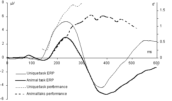
categorization task, target and non-target were equiprobable). As shown in
the figure below, we first observed a 30-40 ms delay for the categorization
compared to the single image detection task both in terms of differential
ERPs (grand average target minus non-target) and behavioral responses (in
d' accuracy measure shifted by -60 ms so that they would align with the ERPs).

We also observed
similar regions of activity for the two differential ERPs. To interpret this
result, we hypothetized that both tasks recruited the same regions of activation
but that the top-down task preseting of this region depended on the task,
so that the unique image detection task was faster than the categorization
one. (This work is under
the process of being published.)
Spectral
analysis using ICA in the categorization task (human EEG)
Application
of ICA to two sessions (2 consecutive days) of the categorization task for
one subject. We found a good correspondance between the two sessions both
for the ICA components and for their synchronization. We also develop a new
type of representation of brain activity "brainmovie" to vizualize the spectral
correlation (coherence) of many ICA component simultaneously. This work has
been published in Neurocomputing, see publications.
Other relevant publications of theThorpe
group on categorization
Thorpe, S., Fize, D., Marlot,
C. (1996) Nature 1996 Jun 6;381(6582):520-2. Pubmed link
Van Rullen, R., Thorpe
S. (2000) J Cogn Neurosci 2001 May 15;13(4):454-61. Pubmed link
Rousellet, G., Fabre-Thorpe,
M., Thorpe, S. (2002). Nat Neurosci 2002 Jul;5(7):629-30. Pubmed link
EEG changes
accompanying volontary regulation of the 12-Hz EEG activity (BCI)
Jonathan Wolpaw
and this team at the Wadsworth center are tranning subject to control the
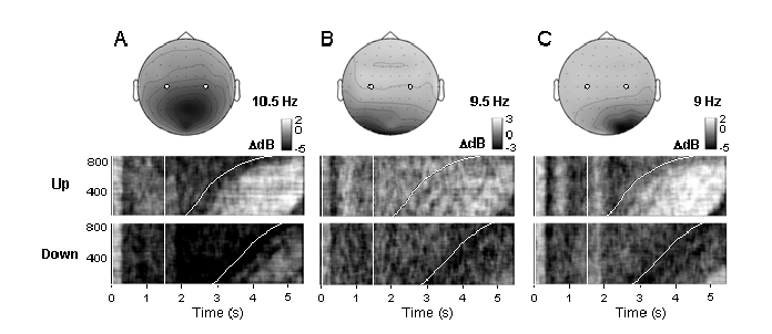
so-called mu rythm at 12 Hz to move a cursor on the screen up and down. We
analsysed some of their data and observed that 12Hz regulation at few electrode
sites are accompanied by large changes at other sites and in other frequency
bands. The figure below shows the behavior of 3 components (A, B, C) at different
frequencies for up-regulated and down-regulated trials. This work is in the process of being
published in IEEE Transactions
on Rehabilitation Engineering. For more detials, see publication.
EEG tools
EEGLAB 4.x
Back in
2000, I wrote a graphical package under Matlab (EEGLAB 2.1) to reject automatically
(or semi-automatically) artifact in EEG data. It was designed to be user
friendly and fully scriptable (all the command can be executed from Matlab
scripts). It was based on the former ICA toolbox package
for Matlab and also provided some functions to automatically visualize
independent components. With Scott
Makeig, in 2002, we then decided to fuse the previous version of EEGLAB (2.1) with the ICA toolbox.
We added more data processing functions and extend the capacities of other
functions. We also focussed on making the function more stable and user friendly. EEGLAB 4.x is available
HERE.
Function to import Neuroscan
data files
These are functions
I programmed to read (neuroscan) EEG, AVG and DAT files under Matlab (NeuroScan EEG file formats). Copy these function
into a directory and launch Matlab on this directory. Type the function
without argument to get the help of how to use it. Note updated versions
of these functions are distributed as part of the EEGLAB toolbox.
Click here to see details of the existing
solutions to read Neuroscan CNT data file.
loadeeg.m : to load Neuroscan
EEG file (optimized for speed)
loadavg.m : to load Neuroscan
AVG files
loaddat.m : to load Neuroscan
DAT text files
ldcnt.m: to load Neuroscan CNT
files (by Andrew James, with additions by myself)
Free software overview
for EEG/ERP under Matlab
Here is an incomplete review
of EEG tools (most of them free). I placed a special focus on Matlab which
is quite convenient to process EEG data.
MATLAB
EEGLAB Toolbox for Electrophysiological Data Analysis
Our toolbox for
ICA and spectral analysis for EEG under Matlab. Allow scripting and most
know spectral and single-trial operations.
FastICA
code for Matlab
ICA decomposition.
An alternative to the runica() function in the EEGLAB toolbox (based on
a different ICA algorithm). Intuitive description of ICA. Not dedicated to EEG.
ERPA visualizing
tool under Linux
Tool to visualize
ERPs under Linux. Convenient to determine the latencies and amplitude of
ERP peaks. However, except for this functionality, the software has few
capacities and it is not free (I have not tested the latest version though).
Magnetic/Electric Source Analysis, User Interface
I did not tested
it. It runs on all platforms.
Stan - software for EEG/ERP processing
Some C-programs
and Neuroscan processing functions under Linux/Windows (average, artifacts,
events...). Under Windows, a primitive graphic interface is also available
(I did not test it).
Brainstorm
EEG (MEG) Source
localization using Matlab. Can map dipole locations onto MRI data. Can
not yet use the scructural MRI properties for modeling though. Also, there
is not scripting language.
Alois' Matlab page
Detailed Matlab
page for EEG (not ERP) file format. Also on this page, the Adaptive Autoregressive
Modeling Matlab toolbox for online processing of EEG (I did not tested it
though).
EEG Toolbox
Tools for looking
at ERPs (some functions of EEGLAB for reading data are also included). The
function for determining the latency of ERP peaks is worth trying. The error
handling is horrible though: the toolbox keeps on generating strange
errors and you don't really know why.
Matlab
and EEG
Review of the
tools available to process EEG data with Matlab.
Mathtools
Mathtools is
a technical computing portal for all scientific and engineering needs.
The portal is free and contains over 20,000 useful links to technical computing,
covering C/C++, Java, Excel, Matlab, Fortran and others.


