The EEGLAB News #14
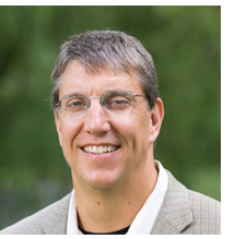 Daniel Ferris, Ph.D.
Robert W. Adenbaum Professor in Engineering Innovation, J. Crayton Pruitt Family Department of Biomedical Engineering, University of Florida
Professor, Department of Mechanical Engineering,
Professor, Department of Neurology, University of Florida
Biomechanics, neural control, locomotion, and robotics
Human Neuromechanics Laboratory at the University of Florida
“A main goal of our lab is to push the limit on the type of physical activity that our subjects can do while we record their electrocortical activity with EEG. Some of our most recent studies have subjects running, jumping obstacles, and playing table tennis, while we study their brain activity.”
Dr. Daniel Ferris, Director of the Human Neuromechanics Laboratory at the University of Florida, is passionate about using Mobile Brain/Body Imaging (MoBI) to study neural control and biomechanics of human locomotion. In other words, he studies how humans walk, run, and move, both in health and disability. “My research interests are a combination of basic science, technology development, and clinical translation around human movement,” he shares.
The techniques and equipment used in his lab sound like a science fiction novel set in a futuristic gym: “We use many techniques,” he describes, “such as mobile brain imaging, lower limb bionic prostheses, and lower limb robotic exoskeletons. Our lab equipment includes a force instrumented treadmill, overground forceplates, high-density electroencephalography (EEG), electromyography (EMG), and a 20 camera motion capture system. Our goals are to develop new technology for assisting with human locomotion, merging human and machine with mechatronic brain-computer interfaces.”
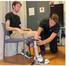 Mobile Brain/Body Imaging (MoBI). Dr. Ferris’ early interest in human movement led him directly into the field of MoBI. In 2002, while a professor at the University of Michigan, Dr. Ferris began studying how humans adapt to walking with robotic exoskeletons. “Our studies showed that some people learned really quickly, some took a long time to learn, and some subjects could be easily disrupted in the learning process by talking with them to distract them,” he recalls. “I wanted to know what was going on in their brains as they were learning how to walk with the robotic exoskeletons. I searched for places to study brain imaging techniques that would allow me to study mobile subjects." (Photo: Dr. Ferris fitting an exoskeleton on a participant; Photo credit: UF)
His search led him to Dr. Scott Makeig and the Swartz Center for Computational Neuroscience (SCCN) at UC San Diego. "I spent six months with Dr. Makeig and his laboratory in 2008, while I was on sabbatical from my faculty position at the University of Michigan," he recollects. While working in Dr. Makeig’s lab, he was introduced to EEGLAB by Dr. Klaus Gramann (Founder of the Berlin Mobile Brain/Body Imaging Lab (BeMoBIL) at the Technical University of Berlin). “I brought home the approach to my own lab in Michigan,” Dr. Ferris shares gratefully. “Over the years, we have developed new hardware and data processing pipelines that we have combined with more standard EEGLAB analysis.”
 After returning to Michigan, Dr. Ferris collaborated with Dr. Makeig on writing a large ONR grant proposal, which was funded. This MoBI work was immediately impressive. As part of this ONR project, the team became the first in the world to demonstrate that high-density EEG and independent component analysis can provide functional imaging of brain activity during human walking and running. (Gwin JT, et al, 2010) "I collaborated with Dr. Makeig and Dr. Gramann," Dr. Ferris adds. "All the data were collected in my Michigan lab but the three of us were co-authors with Joseph Gwin, one of my Ph.D. students."
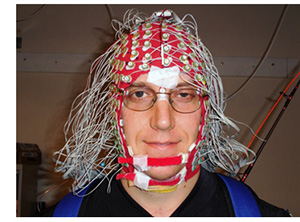
(Photo: Dr. Ferris wearing an EEG cap at SCCN; Photo credit: Dr. Klaus Gramann, a postdoc in Dr. Makeig’s lab at the time).
After returning to Michigan, Dr. Ferris collaborated with Dr. Makeig on writing a large ONR grant proposal, which was funded. This MoBI work was immediately impressive. As part of this ONR project, the team became the first in the world to demonstrate that high-density EEG and independent component analysis can provide functional imaging of brain activity during human walking and running. (Gwin JT, et al, 2010) "I collaborated with Dr. Makeig and Dr. Gramann," Dr. Ferris adds. "All the data were collected in my Michigan lab but the three of us were co-authors with Joseph Gwin, one of my Ph.D. students."
(Photo: Dr. Ferris wearing an EEG cap at SCCN; Photo credit: Dr. Klaus Gramann, a postdoc in Dr. Makeig’s lab at the time).
In 2017, Dr. Ferris relocated from Michigan to the University of Florida. His current lab uses tools from neurophysiology, biomechanics, and engineering to identify general strategies humans use to control gait in health and disability. The long term goal is to create better robotic devices for assisting with human movement. He has developed robotic lower limb exoskeletons for ankle, knee and hip joints, as well as for the whole limb. This research has provided important insight into the most effective ways to add mechanical assistance for locomotion.
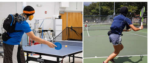 “New technology and devices that allow us to study and assist human movement is where I have the most fun!” Dr. Ferris admits. One example of the fun (and hard work) in Dr. Ferris’ Human Neuromechanics Laboratory is a recent study led by a Ph.D. student, Amanda Studnicki, who also happens to be an avid athlete (and talented tennis player). The study, ‘Characterizing and Removing Artifacts Using Dual-Layer EEG during Table Tennis,’ shows promising results that can benefit future brain science research. The team found that “using noise electrodes for data processing provided cleaner brain components. These results advance technological approaches for recording high fidelity brain dynamics in human behaviors requiring whole body movement.” (Studnicki A, et al., 2022) The team found that “using noise electrodes for data processing provided cleaner brain components. These results advance technological approaches for recording high fidelity brain dynamics in human behaviors requiring whole body movement.” (to view short video clips of the table tennis setup on twitter, click here and here!) (Photos above: Studying brain dynamics during table tennis and tennis; Photo credit: Amanda Studnicki)
 Challenges.
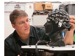
Still, with all of the exciting projects underway, moving mobile brain imaging into more ecologically valid, real world settings is a major challenge for the field. “If we want to understand how the brain functions in everyday life, we need to record its activity in everyday life,” Dr. Ferris explains. “Constrained laboratory tasks and settings will only take us so far.” (Dr. Ferris with electrical phantom head; Photo credit: UF)
Challenges.
Still, with all of the exciting projects underway, moving mobile brain imaging into more ecologically valid, real world settings is a major challenge for the field. “If we want to understand how the brain functions in everyday life, we need to record its activity in everyday life,” Dr. Ferris explains. “Constrained laboratory tasks and settings will only take us so far.” (Dr. Ferris with electrical phantom head; Photo credit: UF)
Another major challenge in the field is coming up with theory to make sense of the new data. “We are able to measure body and brain metrics in novel and multidimensional ways,” Dr. Ferris elucidates. “The overabundance of data means that we need to figure out how to come up with theories that better describe the new data. My personal bias is towards coming up with new ways to merge humans and machines.”
Upbringing. Born in upstate New York, Dr. Ferris spent his first seven years in a small town called North Rose, NY. He then moved to central Florida, where he stayed for his undergraduate degree at the University of Central Florida in mathematics education (1992) and his M.S. degree in exercise physiology at the University of Miami (1994). He then attended UC Berkeley for his Ph.D. in human biodynamics (1998).
His early athletic and vocational experiences helped guide his path. “I was an athlete growing up,” Dr. Ferris explains. “I played football in college and had many injuries. The injuries inspired me to learn more about the function and control of my body.” His undergraduate job at the University of Central Florida library also influenced his future path. “Working at the university library, I checked books in and out that other students and faculty had borrowed,” he recalls. “That led me to read Victor Katch’s textbook on exercise physiology, Eric Kandel’s textbook on neuroscience, and R. McNeill Alexander’s many books on biomechanics.” He remembers being captivated by these topics. “Today,” he laughs, “the research we do in the lab combines all three approaches to studying human movement!”
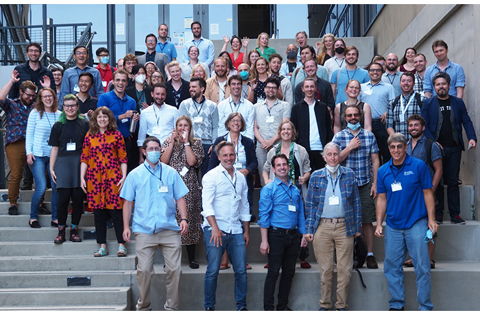
The Future. Dr. Ferris is enthusiastic about the future. “The community of MoBI researchers has grown so much since I joined Scott’s lab in 2008,” he shares, “and I hope it continues to flourish and expand in the next 15 years. We are a supportive and inclusive community that welcomes novices to the field.” (Photo: Attending MoBI 2022 at UC San Diego; Photo credit: Makoto Miyakoshi)
Dr. Ferris is eager to continue exploring the connection between the nervous system and musculoskeletal system, adding: “I want to know how these systems work together to produce the amazing feats of professional athletes, and how they fail in people with disabilities. I would love to develop a very mobile high-density EEG system that lets us record electrocortical dynamics from humans playing a wide range of high physical activity sports like tennis and beach volleyball.” Along with his desire to develop a MoBI system to study human movement in sports, Dr. Ferris also hopes to create ways for people to better work with and understand the data. “I hope to create an automated system for processing and interacting with that data for scientists, clinicians, and coaches."
Dr. Ferris pauses, then concludes with added emphasis, “How can we create a wildly successful new training approach and/or training technology to improve motor function in healthy neurologically people and people with impaired nervous systems? I don’t mean small, incremental advances, but major acceleration of learning in a short time period?” With his dedication and passion, Dr. Ferris is determined to find the answer.
R. Weistrop, November 2022